Tulane University Students Uncover New Anatomical Feature
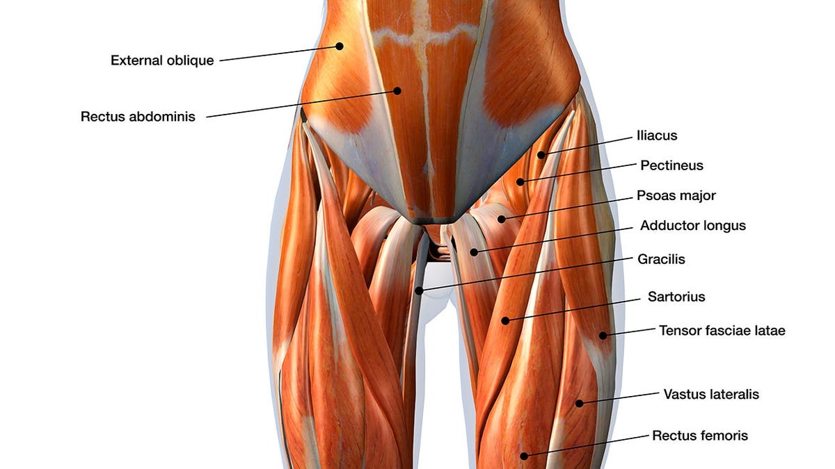
Two undergraduate students at Tulane University unearthed an anatomical feature that even their professor couldn’t initially identify. Dominic Roy and Cooper Bruce, both 19-year-old sophomores at the time, made this significant finding in 2024.
In an interview, Roy recounted an ordinary day volunteering in the cadaver lab alongside R. Shane Tubbs, a professor of neurosurgery and director of surgical anatomy at Tulane School of Medicine. They were involved in dissecting hip specimens to analyze the interconnection between the iliacus and the psoas major, a substantial muscle extending from the lower back through the pelvis to the upper thigh.
Roy remarked, “I noticed a muscle on the other side of the Iliac fascia near the inguinal area of the pelvis.”
This unexpected find intrigued both students and their professor, Dr. Tubbs, as it was not part of their regular discussions. They consulted books and literature but found no previous documentation of the muscle, suggesting a new discovery.
Physicians from overseas, along with budding medical students, also played a role in this discovery. As they examined the specimens and took photos, Roy expressed awe at how swiftly events unfolded.
Their findings, verified and published in the journal Clinical Anatomy, highlighted that Roy and Bruce identified an unexamined part of a muscle deep in the pelvis. This muscle runs to the front of the hip.
Potential Medical Applications
Although the function of the newly discovered muscle remains unclear, experts suspect it may aid in stabilizing the psoas major, involved in hip flexion. The next investigative step is to explore whether MRI scans can visualize the muscle and ascertain if its presence correlates with symptoms.
Dr. Tubbs emphasized the need for further research to understand the muscle’s role. He proposed that awareness of this muscle could benefit patients undergoing minimally invasive procedures and hip surgeries.
Physical therapy might strengthen the muscle if its weakness contributes to symptoms. Alternatively, injections could mitigate pain if overactivity of the muscle is detected.
Surgeons frequently encounter this muscle during hip operations. Understanding its relevance could enhance patient care, particularly if symptoms align with potential muscle-related issues.
Anatomical Variations
This muscle wasn’t present in every examined cadaver, underlining human anatomical variability.
Tubbs commented, “Human beings display significant internal and external variability, and no two individuals are identical.”
The discovery of this muscle in gorillas hints at an evolutionary link. Bruce pondered why this muscle persists in some humans and not others.
The nature of cadaver studies, devoid of personal histories, poses challenges in correlating specific activities or lifestyles with this anatomical feature.
Insights Beyond Textbooks
Exposure to real cadavers provides prospective doctors with invaluable experience, enhancing their understanding beyond synthetic models. Despite advancements, ongoing discoveries in human anatomy continue, even into 2026.
The students’ groundbreaking work may ultimately gain inclusion in anatomical textbooks, inspiring their continued pursuit of medical careers.